
Introduction
- Strains of bacteria which are motile possess filamentous, cytoplasmic appendages protruding through cell wall called flagella.
- This effect screw like propulsive movements and act as organs of locomotion.
- They are much thinner than the flagella or cilia of eukaryotes.
- The flagellum is a long, thin filament, hair-like, un-branched, twisted spirally in an open, regular wave form.
- It is about 0.02 micron thick and is usually several times the length of the bacterial cells.
- It can be seen with the light microscope only after being stained with special stains that increase their diameter.
- Electron microscope is also used to observe flagellum and when stained with phosphotungstic acid it appears as hollow tube.
- It originates in the bacterial protoplasm and is composed entirely of protein called flagellin.
- According to the species, flagella have the characteristics patterns of distribution in the bacterial cell.
- There are four types of flagellar distribution on bacteria. They are:
Types of flagella
1.Monotrichous: single polar flagellum. E.g. Vibrio cholera Campylobacter spp., Caulobacter crescentus, etc.
2.Amphitrichous: single flagellum attached to each end. E.g. Alkaligenes faecalis, Magnetospirillum, etc.
3.Lophotrichous: tufts of flagella at one or both ends. E.g. spirilla, Pseudomonas fluorescens, etc.
4.Peritrichous: numerous flagella all over the bacterial body. E.g. typhoid bacilli, E. coli, Salmonella, Klebsiella, etc.
5.Atrichous: absence of flagella. E.g. Shigella.

Image source: wikipedia
Structure of flagellum
- Each flagellum consists of various components and moves by rotation, much like a propeller of a boat motor.
- It consists of three distinct parts. They are: helical, long filament, short hook and basal body.
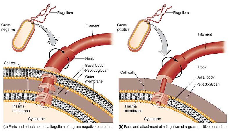
- The filament lies external to the cell and the hook-basal body portion is embedded in the cell envelope.
- The basal body is attached to the cytoplasmic membrane by ring like structures.
- The chemical composition of the basal body is unknown while the hook and filament are composed of protein sub-units (monomers) arranged in a helical fashion called flagellin.
- Flagellin is highly conserved in amino acid sequences in species of bacteria, which suggests that flagellar motility has deep roots within this evolutionary domain.
- The flagella are driven by the rotary action of the swivel-like basal-hook.
- The motor portion is present at the base of filament which is anchored in the cytoplasmic membrane and cell wall.
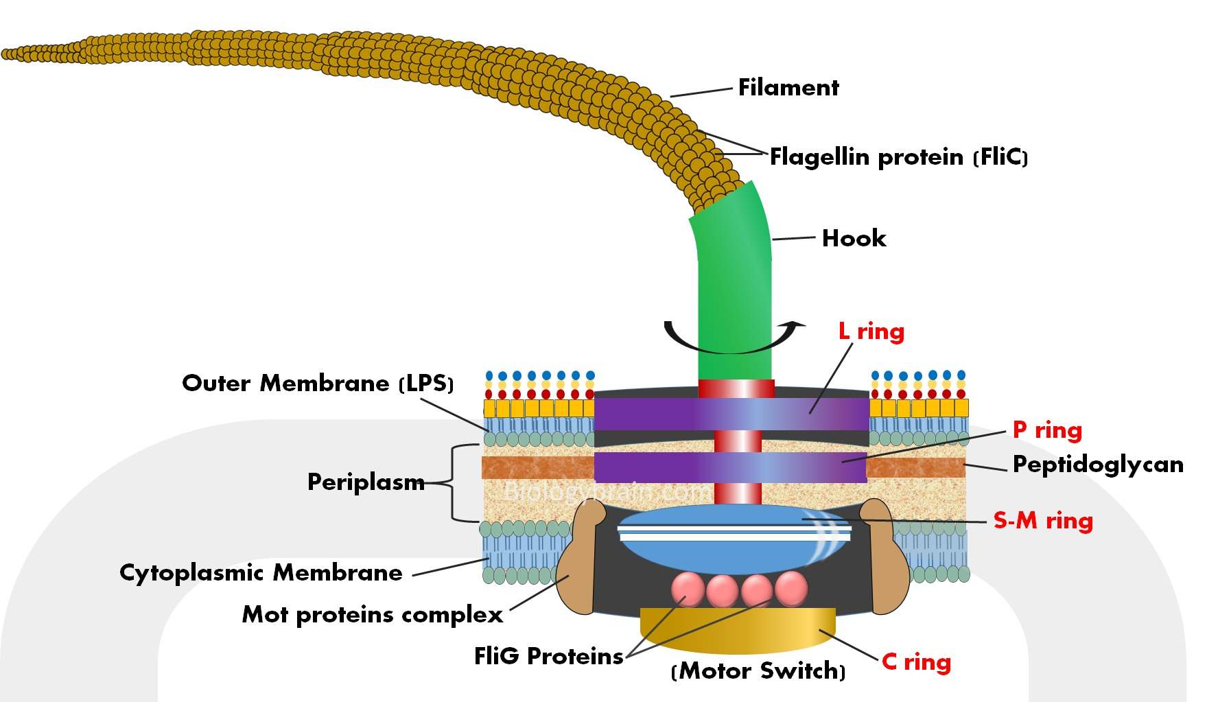
Image source: biologybrain
- The motor has a central rod that passes through a series of rings.
- An outer ring called L-ring is anchored in the lipopolysaccharide layer in gram negative bacteria.
- The second ring called the P-ring is anchored in the peptidoglycan layer of the cell wall.
- MS and C rings, which are the third set of rings, are located within the cytoplasmic membrane and the cytoplasm respectively.
- Only inner pair of rings is present in the case of gram positive bacteria as it lacks an outer membrane.
- Mot proteins are present in series which surrounds the inner ring and are anchored in the cytoplasmic membrane.
- The Fli proteins which are a final set of proteins function as the motor switch.
- These proteins reverse the direction of rotation of flagella in response to intracellular signals.
References:
i) https://www.intechopen.com/chapters/39455
ii) https://microbenotes.com/flagella/