

- Entamoeba histolytica is a common parasite in the large intestine of humans, certain other primates and some other animals.
- This parasite is responsible to cause amoebiasis which is the third leading parasitic cause of death in the developing countries.
- Tropohozoites of this parasite are found in the mucous and sub-mucous layers of the large intestine of man.
Morphology of E. histolytica
The parasite generally occurs in three stages. They are: trophozoite, pre-cyst and cyst.
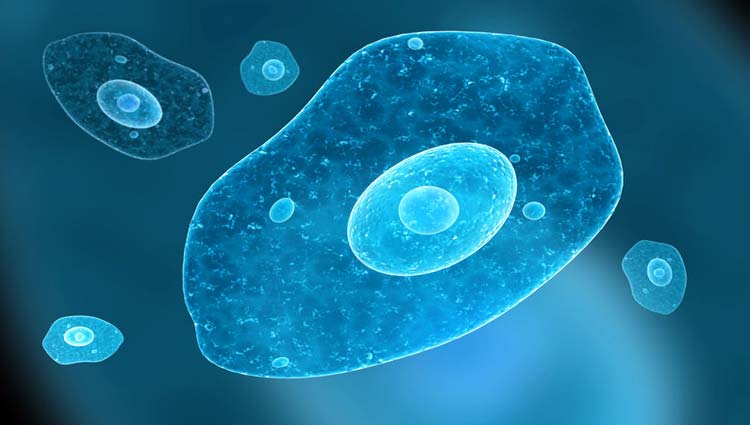
a) Trophozoite ( The Growing or Feeding stage)
- It is the invasive form of the parasite.
- The shape is not fixed because of constantly changing position.
- The size ranges from 18- 40 µm or being 23- 30 µm.

Image source: alamy
- The cytoplasm is divided into two portions; a clear translucent ectoplasm and a granular endoplasm.
- RBCs, occasionally leukocytes and tissue debris are found inside the endoplasm.
- The presence of ingested erythrocytes is the characteristics feature of E. histolytica but not E. dispar.
- Nucleus is single, spherical in shape and varying in size from 4- 6µm.
- It has a fine peripheral chromatin and central karyosome.
- Trophozoites are actively motile with the help of their pseudopodia, which is the clear hyaline cytoplasm.
- These may be long and narrow (finger-like) or short and wide.
- They exhibit gliding movement.
- The trophozoites also have filopodia, usually not seen by light microscope.
- Trophozoite is an anaerobic parasite.
- It nourishes mainly on the bacteria and cytolysed tissue substances.

b)Pre-cyst
- Stage between trophozoite and cyst.
- Smaller in size ranging from 10-20 µm.
- It is round or slightly ovoid with a blunt pseudopodium projecting from the periphery.
- The endoplasm is free of RBCs and other ingested food particles.
- It contains a large glycogen vacuole and two chromatid bars that are rounded at the ends.
c)Cyst (the infective form)
- During encystment, the parasites become rounded and surrounded by a highly refractile membrane called the cyst wall.
- Small, spherical, measures 10 -16µm in diameter.
- Cytoplasm is granular and contains rod-like chromatid bars and glycogen masses but no RBCs or food particles.
- The nucleus shows the same characteristics as that of the trophozoite nucleus.
- The mature cyst contains four nuclei (quadrinucleate cyst).
- The cyst begins as a uninucleate body soon divides by binary fission and develops into binucleate and quadrinucleate bodies.
- During the process of division, the nuclei undergo gradual reduction in size, becoming 2 µm in diameter.
- The mature cysts passed in the stool, do not undergo any further development and remain alive for a few days, if they are not dried or heated.
Distinguishing features of E. histolytica (pathogenic) and E. dispar (non-pathogenic)
- Pathogenic E. histolytica are characterized by their ability to:
- Agglutinate the lectin, concanavalin A.
- Cause caecal ulceration in weaning rats.
- Increased phagocytosis of RBCs.
- Adhere to the apical epithelial cells and
- Produce characteristic iso-enzyme patterns (zymodemes) or starch gel electrophoresis.
- E. dispar is non-pathogenic species.
- The species lacks the capacity to invade tissue and is associated with the asymptomatic carrier state.
Life Cycle
- The life cycle of E. histolytica is simple and is completed in a single host, the man.
- The mature quadrinucleate cysts are the infective forms of the parasite.
- Man acquires infection by ingestion of water and food contaminated with mature quadrinucleate cysts.
- Infection is also acquired directly by ano-genital or oro-genital sexual contact.
- The fully developed cysts gains entrance into the alimentary canal and pass unaltered through the stomach as the cyst wall is resistant to the action of the gastric juice.
- The cyst wall is lysed by the intestinal trypsin.

- Excystation occurs when the cyst reaches the caecum or lower part of the ileum where there is neutral or alkaline medium.
- Each cyst liberates a single amoeba with four nuclei, a tetranucleate amoeba which eventually forms eight amoebulae (metacystic trophozoites) by the division of nuclei with successive fission of cytoplasm.
- These trophozoites are actually motile and carried by peristalsis through the small intestine to the ileo-caecal area of the large intestine.
- Here, they grow and multiply by binary fission.
- They then colonize on the mucosal surfaces and in crypts of the large intestine.
- During growth, E. histolytica secretes a proteolytic enzyme of the nature of histolysin.
- This enzyme brings about destruction and necrosis of tissues and thereby helps the parasite in obtaining nourishment through absorption of these dissolved tissue juices.
- In some individuals, the multiplying trophozoites produce no or little lesion if any in the tissue.
- They feed on the starches and mucus secretions on the surface of mucosa.
- In other individuals, the trophozoites may invade the tissue of the large intestine producing characteristics lesion in the colon through the stages of gelatinous necrosis, abscess and finally ulcer.
- A large number of trophozoites are excreted along with blood and mucus in the stool.
- In a few cases, trophozoites may gain entrance into the liver where they multiply.
- The trophozoites may produce suppurative amoebic liver abscess preceeded by non-suppurtaive infection of the liver.
- A high degree of pathogenicity of the parasite is a disadvantage to itself.
- After sometime, when the effect of the parasite on the host is gradually toned down together with the concomitant increase in the tolerance of the host, the lesions become quiescent and commence to heal.
- The parasite now finds it difficult to continue its life cycle solely as the trophozoite and therefore, prepares to produce strains to survive its extinction.
- A certain number of these trophozoites are discharged into the lumen of the bowel and are transformed into small pre-cystic forms from which cysts are developed.
- The infective cysts can remain viable for months in suitable moist environment.
- These cysts cause infection in other susceptible persons through faecal contamination of water and vegetables or direct faecal oral contact and the cycle is repeated.
References:
i) https://www.ncbi.nlm.nih.gov/pmc/articles/PMC6304615/
ii) https://www.uptodate.com/contents/intestinal-entamoeba-histolytica-amebiasis
iii) https://www.healthline.com/health/amebiasis